Sparing Patients from Surgery with Transcatheter Interventions
From prosthetic heart valve implantation to using radiofrequency energy for pancreatic tumor ablation, skilled specialists are increasingly carrying out high-clinical-impact interventions working over catheters advanced into diseased anatomy from a far-off access site. Achieving a desired therapeutic outcome while avoiding a major surgery can be a huge win for a patient who undergoes a transcatheter intervention. But carrying out a high-stakes intervention while working over a catheter brings tremendous challenges, even for the most capable interventional teams. The limitations of conventional imaging like ultrasound and x-ray can leave teams feeling like they’re flying blind at key junctures during transcatheter procedures.
Ultraslim GlintTM Catheters from Myka Labs: A Revolutionary Advance for Transcatheter Interventions
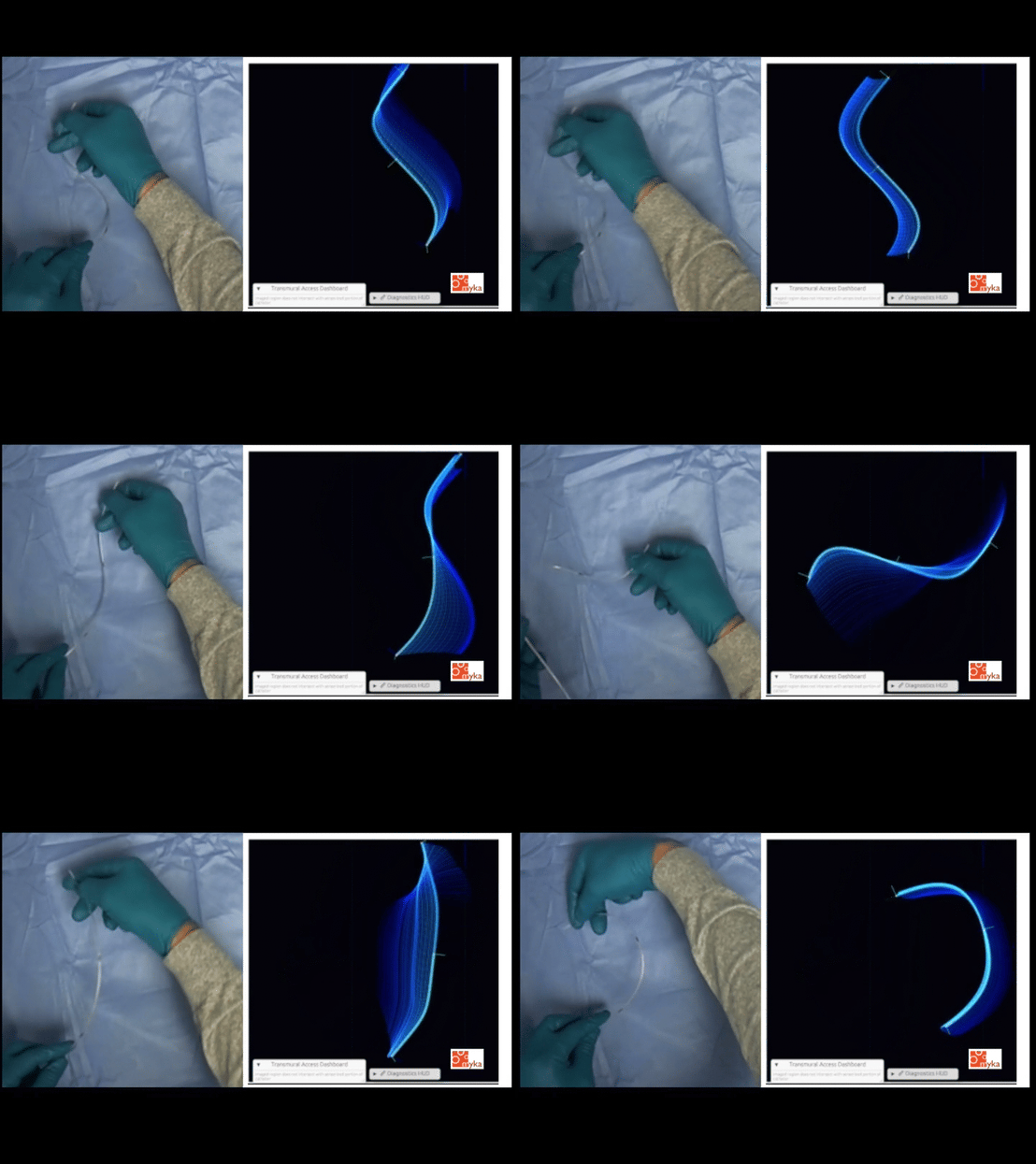
Catheters are widely used for functions like passing contrast and delivering devices—e.g. stents, prosthetic valves—into diseased anatomy. Glint smart catheters from Myka Labs handle basic catheter functionality while also generating a continuous stream of high-value real-time sensor data during procedures. Ultraminiature sensor arrays integrated into the distal end of each Glint smart catheter capture data that is carried up the shaft by thin wires and routed to a bedside processing unit. Glint smart catheter sensor data is the foundation for greater precision, clarity and confidence in high-stakes transcatheter procedures. Critically, Glint smart catheters have ultraslim form factors that allow for straightforward substitution for conventional catheters.
Glint Ultraslim Smart Catheters plus Advanced Artificial Intelligence: A Gamechanging Combination
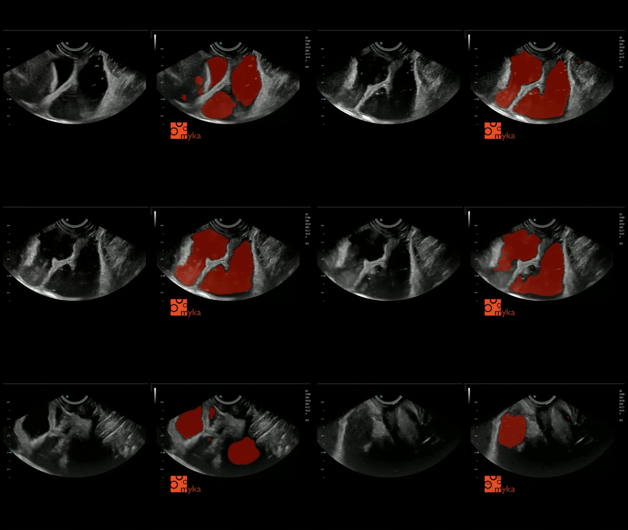
How can data streaming from Glint catheters be used to the greatest effect during high-stakes transcatheter procedures? By using deep learning models to analyze catheter data in combination with conventional intraoperative imaging data feeds. Shown here: imaging slices of abdominal anatomy using a scope-tip-mounted curvilinear ultrasound array operated within the stomach. Myka Labs is at the forefront in developing carefully architected, rigorously trained deep learning models like the Glint real-time vision transformer model that produced the overlays seen here. Blending in catheter data then reveals which of the segmented lumens is the second part of the duodenum, which the jejunum, which the transverse colon–critical insight for achieving procedure objectives.

Endoscopically Repairing Esophageal Atresia using Connect-EA: Favorable Outcomes from Early Clinical Experience
Results of the use of Connect-EA, an advanced esophageal magnetic compression anastomosis (EMCA) system developed at UCSF and that is foundational to Myka’s development of advanced smart catheter technology and accompanying high-performance AI models, have now appeared in multiple peer-reviewed journal articles. Working closely with Dr. Oliver Muensterer of LMU Klinikum, Myka Labs founder and former UCSF pediatric surgeon division chief Dr. Michael Harrison arrived at the design of Connect-EA through exhaustive preclinical studies on the role of compression in minimizing deleterious effects of fibrosis in the post-repair esophagus. Esophageal atresia, a congenital malformation where a portion of the esophagus is missing such that food can not pass from the mouth to the stomach, occurs in roughly one in every 2,500-5,000 live births.